Lentigo
General Information
Lentigo Simplex is the most common form of lentigo. Usually present by birth, but may develop in early childhood as a single lesion or multiple lesions. Lentigo simplex does not occur after sun exposure and it does not have any predisposition of a medical disease or condition. It is also referred to as simple lentigo and juvenile lentigo. Other types of lentigines include Solar Lentigo, which is the most common benign sun induced lesion appearing on the face, arms dorsa of the hands and upper part of the trunk. Ink-spot lentigo limited also to sun-exposed areas of the body, the most common presentation includes one ink-spot among an extensive number of solar lentigines. PUVA Lentigo, which is a pale brown macule, that appears around six months after PUVA therapy for psoriasis. Radiation lentigo, which is considered an indicator of a prior cutaneous exposure to a large single dose of ionizing radiation. Tanning-bed lentigines appearing more commonly on the anterior aspects of the arms and legs, but can also occur on the neck and chest. Oral melanotic macules can appear on the gingiva, buccal mucosa, palate, and tongue; Labial melanotic macules usually occur on the vermilion of the lower lip. Lesions for both are usually solitary, symmetric, and asymptomatic. Vulvar and penile lentigo are benign lesions similar to labial melanotic macules. In men, the most common sites are the glans penis, corona, corona sulcus, and penile shaft. The lesions vary in color from tan to brown to dark brown, and they have irregular borders and skip areas. In women, the lesions appear anywhere on the genital mucosa as a mottled, pigmented patch with skip areas. Lentigo lesions are benign (non-cancerous) but since their appearance is sometimes similar to melanomas or other cancerous lesions they need to be examined carefully by health-care providers.
Epidemiology
Frequency yet to be determined
Etiology
Present at birth or more commonly first develop in early childhood; not induced by sun or associated with any medical diseases
Pathogenesis
Only a few develop initially, but over time up until adult life more may appear
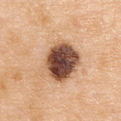
Clinical
Round or oval shaped macule (flat spot or patch) 3-15mm in diameter; edges may be smooth or jagged; color ranging from brown to black
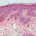
Histology
Slight-to-moderate elongation of the rete ridges with melanocyte proliferation in the basal layer, increased melanin in both the melanocytes and the basal keratinocytes, and the presence of melanophages in the upper dermis.
Bibliography
1. “Lentigo” (Online). June 2007. http://www.emedicine.com/DERM/topic221.htm (visited: March 19, 2008) 2. “Lentigo Simplex” (Online). January 2008. http://www.visualdxhealth.com/adult/lentigoSimplex (visited: March 19, 2008)
Download PDF
![]() Lentigo
Lentigo